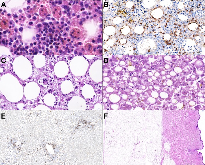
Fig. 4.
scans of histological slides from the autopsy. A – vertebral bone marrow with large macrophages engulfing red cells and lymphocytes (hemophagocytosis). HE, 130x. B – vertebral bone marrow with focal finding of CD8+ lymphocytes rimming the adipocytes; the finding is highly suspect from infiltration by lymphoma, CD8 immunohistochemistry, 55x. C – mesentery with macrophages engulfing lymphocytes, HE, 73x. D – liver tissue showing marked dystrophic changes including steatosis and cholestasis of hepatocytes. HE, 70x. E – liver tissue without apparent lymphoma infiltration. CD3 immunohistochemistry, 12x. F – sample from skin and subcutis tissue without apparent lymphoma infiltration. HE, 2,5x

