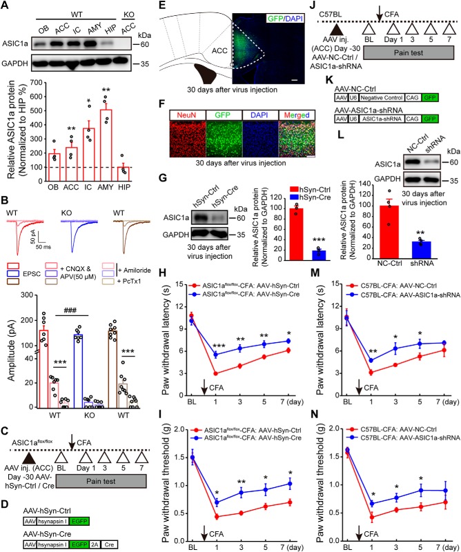
Figure 1.
Genetic manipulations of ASIC1a in ACC influence inflammatory pain hypersensitivity. A, Representative images and quantification of Western blots showing ASIC1a protein enrichment in ACC and several other brain regions. Data represent optical density values normalized to that of hippocampus. n = 4 mice. OB, Olfactory bulb; IC, insular cortex; AMY, amygdala; HIP, hippocampus. B, Representative traces of EPSCs recorded with a high basal stimulation intensity before and after APV (50 μm) + CNQX, and after amiloride or PcTx1 in WT or ASIC1a KO mice. Bottom, Quantification of the ASIC-mediated synaptic currents. n = 7 or 8 neurons/3 mice for WT; n = 7 neurons/4 mice for ASIC1a KO. C, Schematic diagram of experimental procedure for conditional deletion experiments. D, Schematic illustration of the viral vectors for AAV-hSyn-Ctrl and AAV-hSyn-Cre. E, Confocal image showing efficient expression of AAV-hSyn-Cre in ACC of an ASIC1aflox/flox mouse. Scale bar, 200 μm. F, Colabeling of virus-derived GFP with the neuronal marker NeuN. Scale bar, 100 μm. G, Representative images and quantification of Western blots showing that AAV-hSyn-Cre injection reduced ASIC1a expression in ACC (n = 3 mice per group). H, I, ACC-specific deletion of ASIC1a significantly attenuated CFA-evoked thermal hyperalgesia (H) and mechanical allodynia (I). n = 8 mice for each group. J, Schematic diagram of experimental procedure for knockdown experiments. K, Schematic illustration of the viral vectors for AAV-NC-Ctrl and AAV-ASIC1a-shRNA. L, Representative images and quantification of Western blots showing that AAV-ASIC1a-shRNA injection reduced ASIC1a expression in ACC (n = 4 mice per group). M, N, Genetic knockdown of ASIC1a in ACC produced significant analgesic effects in thermal (M) and mechanical (N) pain tests. n = 8 mice per group. BL, Baseline; Ctrl, control. Data are mean ± SEM. *p < 0.05, **p < 0.01, ***p < 0.001, ###p < 0.001.

