Figure 3.
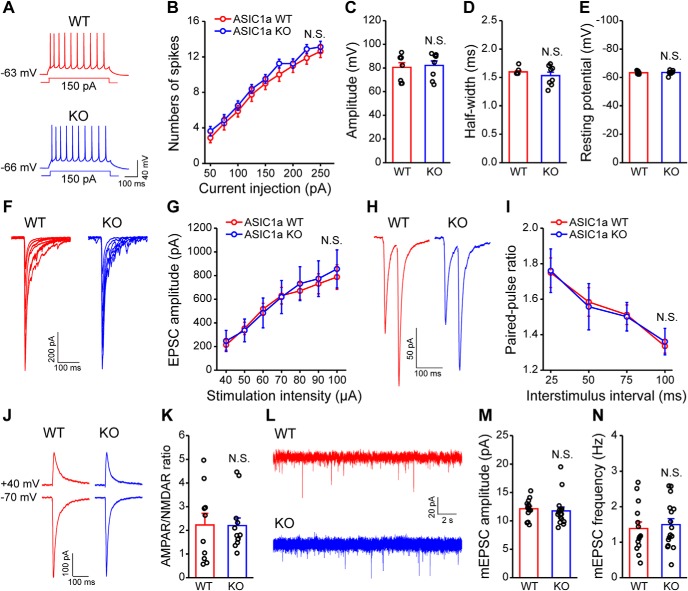
Intrinsic membrane properties and baseline synaptic transmission are unaltered in ACC of ASIC1a KO mice. A–E, Representative traces (A) and pooled data (B–E) showing no effect of ASIC1a deletion on action potential firing properties in ACC pyramidal neurons. B, Intensity-dependent increase in number of spikes elicited in ACC. C, Action potential amplitude. D, Action potential half-width. E, Resting membrane potential of ACC neurons in ASIC1a WT and KO mice (n = 8 neurons/3 or 4 mice). F, G, Representative traces (F) and quantification of evoked EPSCs (G) showing no effect of ASIC1a deletion on input-output relationship of excitatory synaptic transmission in ACC (n = 8 neurons/4 mice). H, I, Representative plots (H) and quantification (I) of paired-pulse ratio recordings showing no effect of ASIC1a deletion on the probability of presynaptic neurotransmitter release (n = 9–11 neurons/4 or 5 mice). J, K, Representative traces (J) and quantification (K) of AMPAR/NMDAR ratios in ACC demonstrating no effect of ASIC1a deletion (n = 10–12 neurons/3 mice). L–N, Representative traces (L) and quantification (M,N) revealing no obvious difference in the amplitude (M) or frequency (N) of mEPSCs recorded in ACC pyramidal neurons (n = 14–16 neurons/4 or 5 mice). Data are mean ± SEM. N.S., not significant.

