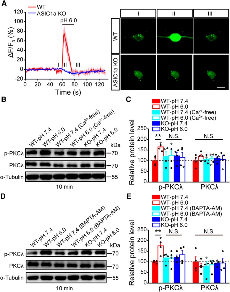
Figure 9.
ASIC1a induces PKCλ phosphorylation through intracellular Ca2+ increase. A, Acid (pH 6.0)-induced changes in cytosolic Ca2+ signal, indicated by GCaMP6 fluorescence, in cultured WT or ASIC1a KO mouse cortical neurons. n = 9 or 10. Thick lines indicate mean. Shaded areas represent SEM. Right, Representative images of GCaMP6-labeled neurons at indicated time points (I, II, and III). Scale bar, 10 μm. B, C, Representative images (B) and quantification (C) of Western blots showing that the acidic solution, pH 6.0, treatment elicited a significant increase in the phosphorylation of PKCλ in cultured cortical neurons from WT but not ASIC1a KO mice. A Ca2+-free pH 6.0 solution failed to evoke PKCλ phosphorylation. n = 4. D, E, Representative images (D) and quantification (E) of Western blots showing that chelating intracellular Ca2+ by BAPTA-AM also eliminated the acidic solution-induced PKC phosphorylation. n = 4. **p < 0.01, N.S., not significant.

