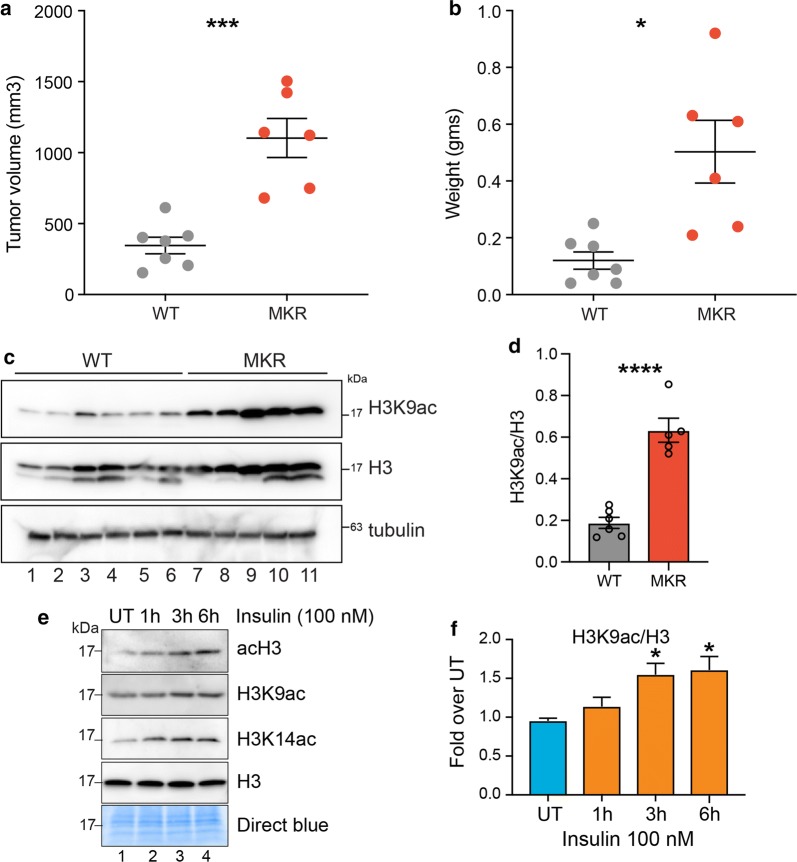
Fig. 1.
Hyperinsulinemia in MKR mice and in vitro promotes an increase in histone acetylation. a Tumor volume (mm3) and b weight are shown for tumors from Rag/WT (WT) and Rag/MKR (MKR) mice. Significance was calculated using unpaired Student’s t test. *p < 0.05, ***p < 0.001. c Western blot analysis showing the total levels of H3K9ac and H3, tubulin controls, in WT and MKR tumors. d Densitometric quantification of H3K9ac/H3 western blot signals in c. Values are Mean + SEM; n = 6 for Rag/WT and n = 5 for Rag/MKR lysates. Significance was calculated using unpaired Student’s t test. ****p < 0.0001. e Western blot analysis using the indicated antibodies in MDA-MB-231 (TNBC cell line) cell lysates treated with insulin (100 nM) for 1 h, 3 h or 6 h. f Densitometric quantification of H3K9ac/H3 western blot signals in (e). Data are represented as fold change over UT. Values are Mean + SEM; n = 3. Statistical significance was calculated using one-way ANOVA, Dunnett’s multiple comparisons test. *p < 0.05

