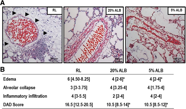
Fig. 3.
Diffuse alveolar damage (DAD) score. a: Representative photomicrographs (light microscopy) of lung parenchyma stained with hematoxylin and eosin. Photomicrographs are representative of data obtained from lung sections of 6 animals (original magnification, × 200). Arrows indicate perivascular edema. Bars = 200 μm. b: Data are shown as median [interquartile range]. The Kruskal–Wallis test was used to compare groups. * vs. RL (p < 0.05)

