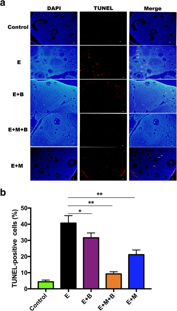
Fig. 6.

The effect of chemotherapy and different treatments on ovarian cell apoptosis. a Ovarian cell apoptosis was analyzed by TUNEL staining. Blue fluorescence indicated cell nucleus stained by 4′, 6-diamidino-2-phenylindole (DAPI). TUNEL-positive cells labeled red (Alexa Fluor 640) indicated cell apoptosis (white arrow). × 200 magnification. Scale bars: 50 μm. b Quantitative analysis showing the percentage of TUNEL-positive cells in each group (n = 3 per group). *P < 0.05 and **P < 0.01 v.s. E group
