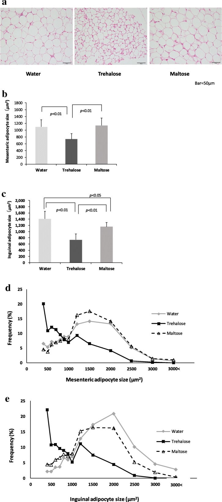
Fig. 5.

The effect of drinking water containing trehalose on adipocyte size in mesenteric and inguinal adipose tissue. The histology of WAT in the mesenteric and the inguinal adipose tissues and also the adipocyte size of both WATs were measured using the image software cellSens. Representative images of hematoxylin-eosin staining in sections of inguinal adipose tissue (× 400) are shown to assess histologically (a) and determine the size of mesenteric adipocytes (b) and inguinal adipocytes (c). In addition, cell size profiling in mesenteric adipose tissue (d) and inguinal adipose tissue (e) is summarized. Values are shown as means ± standard deviations (n = 8). Statistical analysis was performed with Tukey-Kramer. Values show statistical significance (5b and 5c; p < 0.01, 5c; p < 0.05). WAT: white adipose tissue
