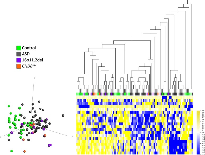
Fig. 1.
Whole blood DNAm comparison between heterogeneous ASD group (n = 52) and age-, sex-matched controls (n = 30). Following filtering by variance (40,550 sites), limma regression and Mann-Whitney U comparison, no CpG sites meet significance criteria of q ≤ 0.05. The 22 sites shown at uncorrected p < 0.001, |Δβ| ≥ 5% do not distinguish clearly between ASD cases and controls. Hierarchical clustering (Euclidian) and principal component analysis (PCA, first 3 principal components labeled) plot show that 16p11.2del (purple) and CHD8+/− (orange) cases are mixed with both heterogeneous ASD cases (gray) and controls (green). Data are normalized for visualization (mean = 0, variance = 1). In heat map, yellow represents high methylation and blue represents low methylation

