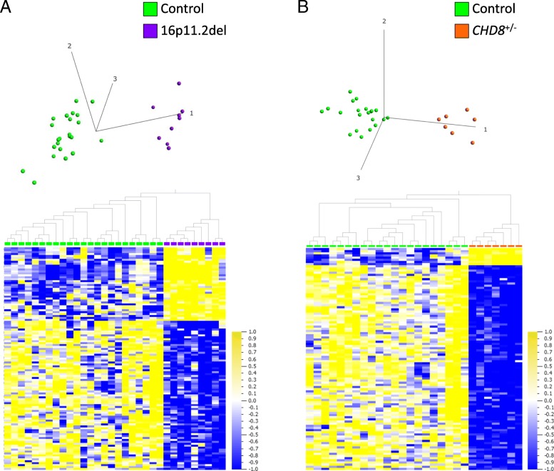
Fig. 2.
a, b DNAm signatures identified in whole blood of individuals with 16p11.2del (600 kb risk locus) or CHD8+/−. a Hierarchical clustering and PCA plot (first 3 principal components labeled) show that 16p11.2del training cases (purple; n = 9) are distinct from age-, sex-matched controls (green; n = 23) at the DNAm signature sites (115 CpG sites; q < 0.05, absolute methylation difference (|Δβ| ≥ 5%). b Hierarchical clustering and PCA plot (first 3 principal components labeled) show that CHD8+/− training cases (orange; n = 7) are distinct from age-, sex-matched controls (green; n = 21) at DNAm signature sites (103 CpG sites; q < 0.01, |Δβ| ≥ 5%) used for classification. Only a single CpG site overlaps between the 16p11.2del and CHD8+/− DNAm signatures: cg25970491 (CLTCL1), which is hypomethylated in both CHD8+/− and 16p11.2del cases relative to controls. All data are normalized for visualization (mean = 0, variance = 1). In heat map, yellow represents high methylation and blue represents low methylation

