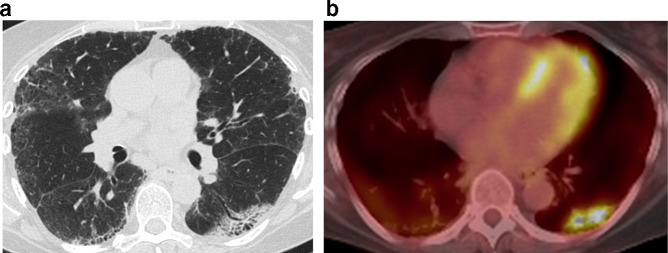
Figure 12.
An irregular mass-like opacity is present in the left lower lobe in a region of fibrotic parenchyma (a). Note a mild background of subpleural fibrosis in the remaining lobes. Fluorodeoxyglucose avidity was apparent on positron emission tomography/CT (b). Histology revealed squamous cell carcinoma.

