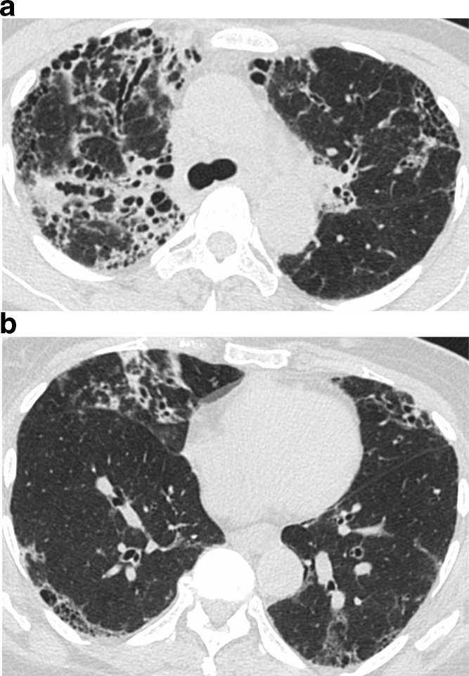
Figure 5.
HRCT features suggestive of an alternative diagnosis. There is worse fibrosis on the cranial image (a) compared to a more caudal level (b), with traction bronchiectasis and distortion showing subpleural and bronchovascular distribution. The upper lung predominance suggests a non-IPF diagnosis. This patient was diagnosed with sarcoidosis. HRCT, high-resolution CT; IPF, idiopathic pulmonary fibrosis.

