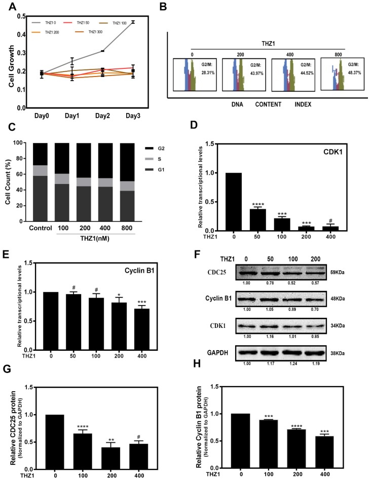
Figure 1.
THZ1 inhibited proliferation of LECs through G2/M phase cell cycle arrest. (A)The portion of viable LECs was determined by MTT assay after treatment of THZ1 (0, 50, 100, 200 and 400nM) for 24 h, 48 h and 72 h. (B) LECs were subjected to 0, 100, 200, 400, 800nM of THZ1. Flow cytometry results of cell cycle phase are shown. (C) Bar graphs represent the mean ± S.E.M. of three independent experiments. (D, E) The mRNA expression level of CDK1 and Cyclin B1 were detected by RT-PCR after treated by various concentrations of THZ1 for 24 h. (F) The protein expression levels of CDC25, Cyclin B1, CDK1 were detected by western blot analysis. Quantification of protein level showed in the G and H. Results are presented as the mean ± SD from three independent experiments. *P < 0.05, **P < 0.01, ***P < 0.001 and ****P < 0.0001 vs. Control.

