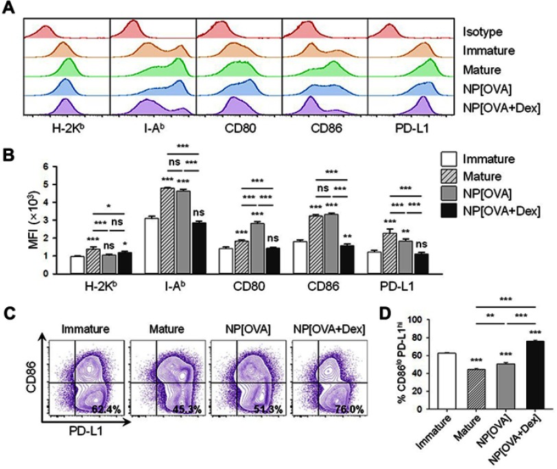
Figure 1.
Effects of NP[OVA+Dex] on the expression of cell surface molecules in DCs.
Notes: (A) DCs generated from the bone marrow cells of C57BL/6 mice were stimulated with IFN-γ (50 ng/mL) plus TNF-α (50 ng/mL), or treated with NP[OVA] or NP[OVA+Dex] (10 μg/mL as OVA) for 48 h. DCs were stained for CD11c, H-2Kb, I-Ab, CD80, CD86, and PD-L1. CD11c+ cells were gated and analyzed for the expression of cell surface molecules. The data shown are representative histograms of four independent experiments. (B) Mean fluorescence intensities of NP-treated, or untreated DCs. The data are presented as the mean ± SD of four independent experiments. (C) Expression of CD86 and PD-L1 in NP-treated, or untreated DCs. The data shown are representative histograms of four independent experiments. (D) The proportion of CD86loPD-L1hi cells in each experimental group is shown. The data are presented as the mean ± SD of four independent experiments. The significance of the data was evaluated using a Tukey test of one-way ANOVA test. *P<0.05, **P<0.01, ***P<0.001. “ns” indicates no significant difference.
Abbreviations: DCs, dendritic cells; Dex, dexamethasone; IFN-γ, interferon-γ; NP, nanoparticle; NP[OVA+Dex], nanoparticles containing ovalbumin and dexamethasone; NP[OVA], nanoparticles containing only ovalbumin; OVA, ovalbumin; PD-L1, programmed death-ligand 1; SD, standard deviation; TNF-α, tumor necrosis factor-α.

