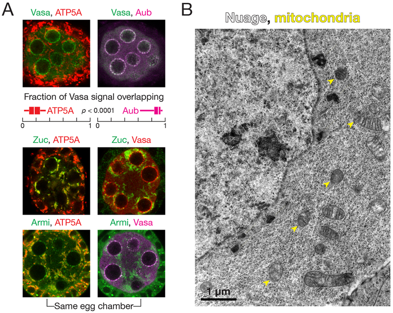
Figure 1. Armi localizes to both nuage and mitochondria, physically separate sites of piRNA biogenesis.

(A) Immunofluorescence detection of Vasa, ATP5A and Aub in wild-type stage 3 egg chambers. Box plots show the fraction of Vasa signal overlapping with ATP5A or Aub. Box comprises the 25th percentile and the 75th percentile of data, with the median indicated; whiskers mark 1.5 × interquartile range. p-value was calculated using the two-tailed Wilcoxon matched-pairs signed rank test. Immunofluorescence of Zuc-3×FLAG or Armi to detect their colocalization with ATP5A or Vasa in wild-type stage 3 egg chambers.
(B) Transmission electron microscopy image of a wild-type stage 3 egg chamber.
See also Figure S1.
