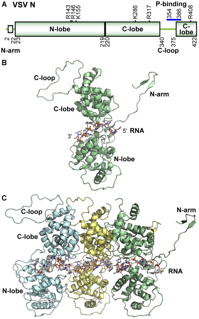
FIGURE 3.

Structure of the VSV N protein. (A) The domain organization of the VSV N protein is schematically represented. Basic residues contributing to RNA binding are noted above the schematic, and residues involved in P-binding are noted with a blue bar. (B) A cartoon representation of the monomeric N protein (PDB id: 2GIC) is shown with bound nine-mer of RNA encapsidated and regional landmarks noted. (C) Assembled trimer of N proteins (each represented in a different color) with bound 27-mer of RNA is shown. All illustrations were prepared with PyMOL (DeLano, 2002).
