
FIGURE 4.
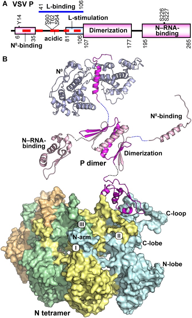
Structure of the VSV P protein. (A) The domain organization of the VSV P protein is schematically represented. Domains are labeled according to known binding partners and function. Six phosphorylation sites are noted above the schematic. (B) VSV P exists as a dimer and is represented in cartoon form with regional aspects noted in (A) labeled. P binds the unassembled N0 (top) via a single helix and adjacent amino acids in the N-terminal intrinsically disordered region (PDB id: 3PMK). The dimerization domain (PDB id: 2FQM) is shown central to the figure. The C-terminal domain of P (PDB id: 3HHZ) binds a bipartite binding site involving the C-loops and an α-helix in the C-lobe of N. A tetramer of N proteins (each represented in a different color) is shown in surface representation. The view is 180 degrees from that in Figure 3C. The three contacts that generate the nucleocapsid are noted: the interactions between (I) the N-arm and the C-lobe on the proximal surface of the left neighboring subunit, (II) the C-loop and the C-lobe of the neighboring subunit to the right, and (III) the N-arm and the C-loop of the N protein subunit two units away on the left.