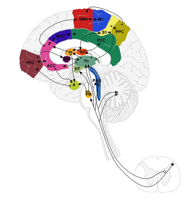
Figure 1.

Schematic representation of Brain Connectome and the Pain Matrix (adapted from Apkarian 2005 and Price 2000 [39,40]). Cortical and sub-cortical pain networks and pathways involved in pain perception. Locations of brain regions involved in pain perception are shown in a schematic drawing showing the regions, their inter-connectivity, and afferent pathways. There are 15 areas in this matrix: anterior cingulate (ACC), medial cingulate (MCC), insula (In), thalamus (Th), prefrontal cortex (PFC), primary and secondary somatosensory cortices (S1, S2), primary and supplementary motor cortices (M1 and SMA), posterior parietal cortex (PPC), posterior cingulate (PCC), basal ganglia (BG), hypothalamus (HT), amygdala (A), parabrachial nuclei (PB), and periaqueductal gray (PAG).
