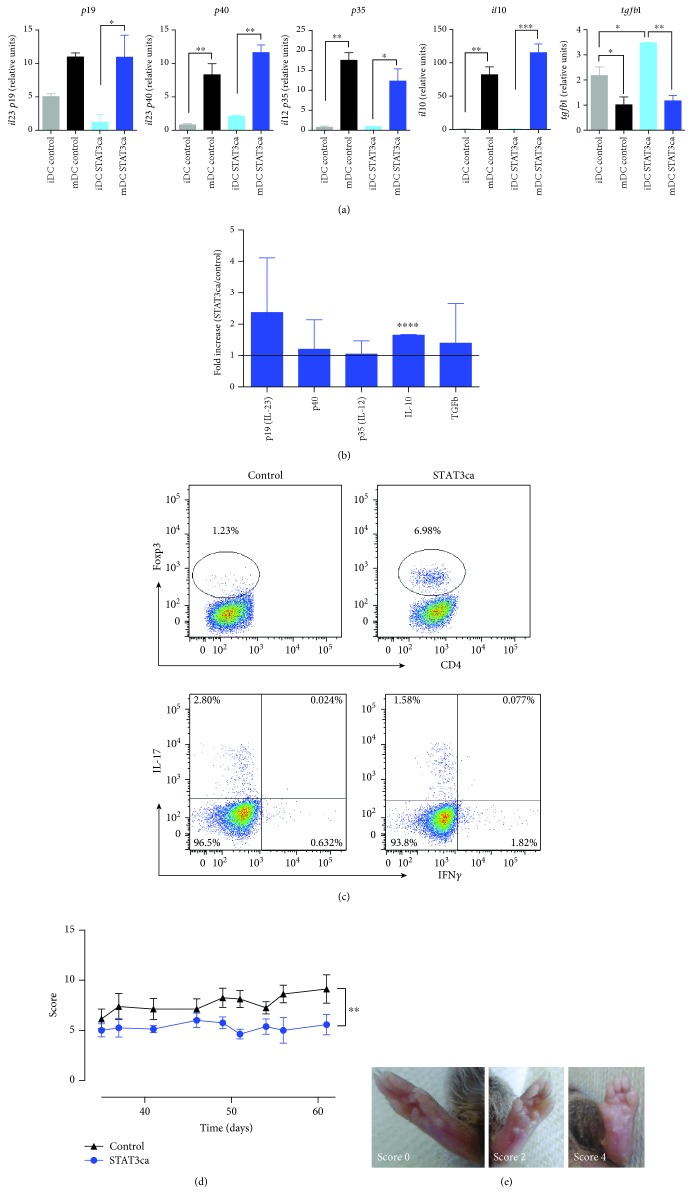
Figure 1.
Permanent activation of STAT3 in DCs induces a tolerogenic profile with therapeutic effects in CIA. Bone marrow-derived precursors from DBA/1J mice were cultured in conditions to differentiate to DCs and transduced on days 4 and 5 with lentiviral vectors (MOI 10) encoding for STAT3ca or with empty pLVX vectors (control) in the presence of polybrene. At day 6, DCs were either left without treatment (iDC) or treated with 500 ng/ml LPS for 24 h (mDC). (a, b) Levels of mRNA encoding for cytokines were evaluated in transduced DCs by quantitative real-time RT-PCR and normalized with the levels of gapdh mRNA. (a) Levels of mRNA encoding for different cytokines are represented as relative units. Values are mean ± SD from three or more independent experiments carried out in duplicate. ∗p < 0.05, ∗∗p < 0.01, and ∗∗∗p < 0.001 by one-way ANOVA followed by Tukey's post hoc test. (b) Data presented as the fold increase of mRNA levels in mDCs relative to mDCs transduced with control vectors. Values are mean ± SD from three or more independent experiments carried out in duplicate. Ratio = 1, which indicates no differences between cytokine mRNA levels in mDCs transduced with STAT3ca and in mDCs transduced with control vectors, is represented by a dotted line. ∗∗∗∗p < 0.0001 by unpaired two-tailed Student's t-test. (c) After lentiviral transduction, DCs were cocultured with naive CD4+ T-cells (at DC : T-cell ratio of 1 : 5) in the presence of anti-CD3ε antibody and incubated for 5 d. Afterward, the percentage of Treg cells was evaluated by intracellular immunostaining of Foxp3 in the CD4+ T-cell population. To determine the percentage of Th1 and Th17 lymphocytes, T lymphocytes were restimulated with PMA and ionomycin in the presence of brefeldin A for the last 4 h of culture and the extent of IFN-γ and IL-17 was assessed by intracellular cytokine immunostaining and analysed by flow cytometry in the CD4+-gated population. Dot plots of Foxp3 versus CD4 (top panels) and IFN-γ versus IL-17 in the CD4+ population (bottom panels) are shown. Representative data from three independent experiments is shown. The percentage of cells present in each quadrant is indicated. (d) After transduction, mDCs were loaded with 40 μg/ml of CII for 18 h and then intravenously administered (5 × 10⁶ transduced DCs/mouse) to DBA/1J mice at day 35 after CIA induction. The extent of disease severity was evaluated along with the disease development as a score described in Materials and Methods. Data represents mean ± SD from eight animals per group. ∗∗p < 0.01 by Mann–Whitney U test. (e) Representative photos of mouse paws displaying no inflammation (score 0, left image), mild inflammation (score 2, middle image), and strong inflammation (score 4, right image).

