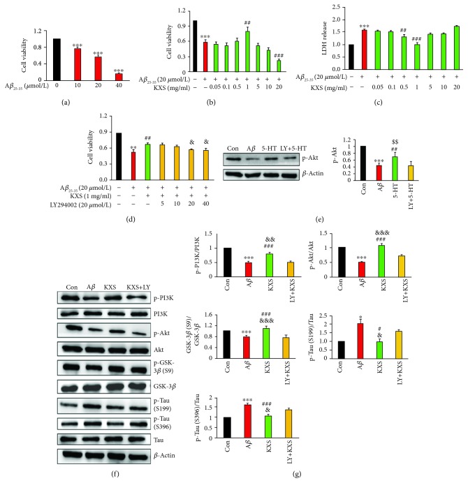
Figure 6.
KXS treatment regulated the activity of neurons and the expression of the Tau signaling pathway in vitro. The treatment of PC12 cells with Aβ 25-35 (10, 20, and 40 μmol/L) alone for 24 h (a) or pretreatment with different concentrations of KXS for 24 h (b) was represented by the MTT assay. The concentration of LDH was assessed as the cytotoxicity assay indicator to assess the effect of different concentrations of KXS (c). The inhibiting effects of LY294002 (5, 10, 20, and 40 μmol/L) were investigated through pretreatment for 1 h (d). The expression of p-Akt was significantly increased by 5-HT (e). KXS had neuroprotection by modulating the hyperphosphorylation of Tau through the PI3K/Akt signaling pathway. Representative western images (f) and quantification (g) of proteins related to the Tau signaling pathway in PC12 cells were represented. Values were the mean ± SD (n = 6). ∗ P < 0.05, ∗∗ P < 0.01, and ∗∗∗ P < 0.001vs. the control group; # P < 0.05, ## P < 0.01, and ### P < 0.001vs. the Aβ group; & P < 0.05, && P < 0.01, and &&& P < 0.001vs. the LY+KXS group; and $$ P < 0.01vs. the LY+5-HT group.

