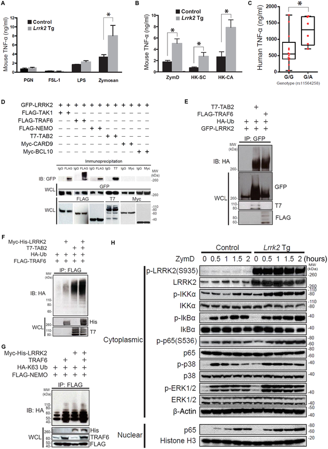
Fig. 2. LRRK2 positively regulates Dectin-1 signaling and interacts with the TAK1 complex.
(A and B) BMDCs from Lrrk2 Tg and control mice were stimulated for 24 hours with the indicated ligands in vitro. The amount of TNF-α in the culture supernatant was measured by enzyme-linked immunosorbent assay (ELISA). (A) Lrrk2 Tg mice, n = 3; control mice, n = 3, P = 0.0263; (B) Lrrk2 Tg mice, n = 3; control mice, n = 3; P = 0.0179 (ZymD), P = 0.0431 [heat-killed S. cerevisiae (HKSC)], and P = 0.0327 [heat-killed Candida albicans (HK-CA)]. (C) DCs from patients with CD (with the G/G or G/A genotype at SNP rs11564258) were stimulated for 24 hours with ZymD in vitro. The production of TNF-α in the culture super-natant was measured by ELISA (G/G genotype, n = 13; G/A genotype, n = 6, P = 0.026). (D) Whole-cell lysates (WCLs) of HEK293T cells cotransfected with the indicated plasmids were subjected to immunoprecipitation with anti- FLAG antibody followed by immunoblotting (IB) with anti– green fluorescent protein (GFP) antibody. IgG, immunoglobulin G. (E) Whole-cell lysates of HEK293T cells cotransfected with the indicated plasmids were subjected to immunoprecipitation (IP) with anti- GFP anti body, followed by immunoblotting with anti- hemagglutinin (HA) antibody. (F and G) Whole-cell lysates of HEK293T cells cotransfected with the indicated plasmids were subjected to immunoprecipitation with anti- FLAG antibody, followed by immunoblotting with anti- HA antibody. (H) Nuclear or cytoplasmic lysates of BMDCs from Lrrk2 Tg mice and littermate control mice were stimulated with ZymD in culture and then were subjected to immunoblotting with antibodies specific to the indicated ligands. Each of the studies is representative of at least three replicates. *P < 0.05 was considered a statistically significant difference. Statistical significance was determined with a Student’s t test. LPS, lipopolysaccharide; PGN, peptidoglycan; IKKα, IκB kinase α; MW, molecular weight.

