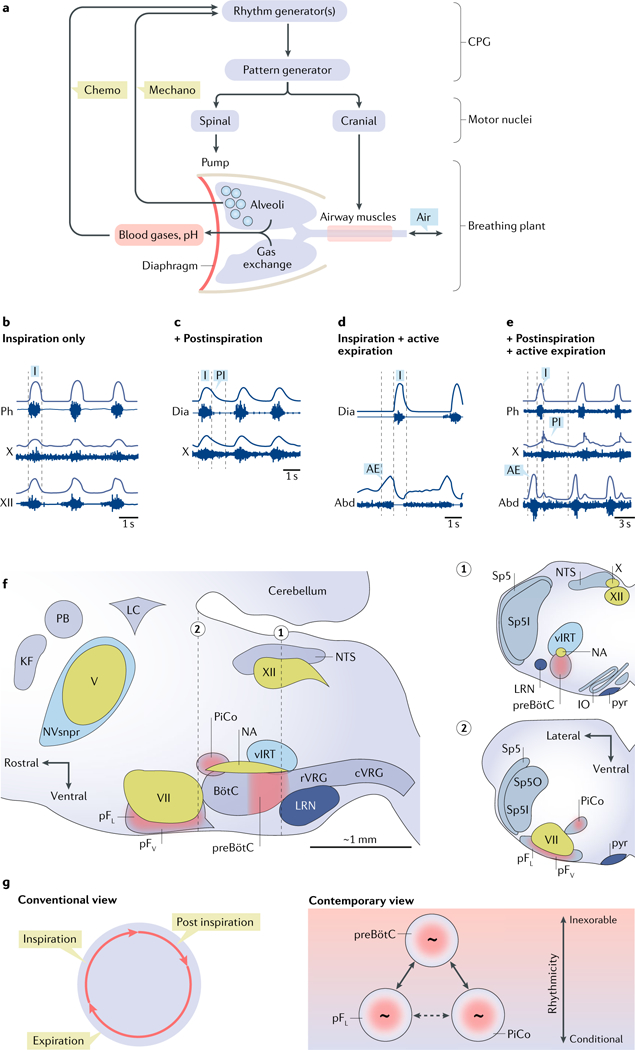
Fig. 2 |. Elements of the breathing central pattern generator. a.

a | Schematic view of breathing neural and physical plant. The image shows a central pattern generator (CPG) composed of rhythm-generating and pattern-generating microcircuits. Spinal and cranial motor nuclei innervate pump and airway muscles, respectively. Airways, lungs, blood gases and sensory (‘chemo’ and ‘mechano’) feedback are indicated. b | Inspiration (I) is the inexorable phase of the breathing cycle. In an anaesthetized rat, the inspiratory cycle is evident in electrical recordings (raw and integrated signals) from the phrenic nerve (Ph), a branch of the vagus nerve (cranial nerve (CN) X) innervating laryngeal adductor muscles, and the hypoglossal nerve (CN XII) innervating the genioglossus muscle (tongue protruder). c | Also in an anesthetized rat, inspiration recorded via diaphragmatic (Dia) electromyography (EMG), shown with postinspiration (PI) recorded from the laryngeal branch of CN X. d | Also in an anesthetized rat, inspiration shown via diaphragmatic EMG (Dia), with active expiration (AE; evoked by disinhibition of the lateral parafacial respiratory group (pFL)) indicated by abdominal EMG (Abd). e | In a reduced in situ rat preparation, inspiration, postinspiration and active expiration measured via electrical recordings from the phrenic (Ph) nerve, CN X and lumbar abdominal (Abd) nerve, respectively. Active expiration was evoked by hypercapnia. f | Parasagittal view of the brainstem containing the breathing CPG. Respiratory rhythmogenic sites are shown in red: the preBötzinger Complex (preBötC; inspiratory), the pFL (expiratory) and the more medial chemosensitive ventral parafacial respiratory group (pFV; rhythmogenic in the perinatal period only), as well as the ‘postinspiratory comp1ex’(PiCo; hypothesized to underlie postinspiration). Other rhythmogenic sites, such as the whisking-related ventral intermediate reticular formation (vIRT) and the masticatory trigeminal principal sensory nucleus (NVsnpr), are shown in blue. Cranial motor nuclei controlling airway resistance muscles, the hypoglossal motor nucleus (XII) and the nucleus ambiguus (NA), as well as facial muscles, the facial motor nucleus (VII) and the trigeminal motor nucleus (V), are shown in green. Brainstem sites associated with breathing motor pattern or sensorimotor integration are shown in grey: the rostral ventral respiratory group (rVRG) containing inspiratory, that is, phrenic and external intercostal, premotor neurons and the caudal ventral respiratory group (rVRG) containing expiratory premotor neurons. Other sites include the pontine Kölliker-Fuse nucleus (KF) and parabrachial nucleus (PB), the nucleus of the solitary tract (NTS) and the expiratory BotC. Also shown is the locus coeruleus (LC) that receives preBötC projections, the cerebellum and the lateral reticular nucleus (LRN). Insets 1 and 2 show transverse sections at the level of the preBötC (dotted line 1) and pF (dotted line 2). Additional structures in the insets include the spinal trigeminal tract (Sp5), the spinal trigeminal sensory nucleus oralis (Sp5O) and the interpolaris (Sp5I), the inferior olive (IO) and the pyramidal tract (pyr). g | In the conventional view of breathing, inspiration, postinspiration and expiration constitute a continuous unitary breathing cycle. According to the contemporary view, inspiration is the inexorable part of the breathing cycle, whereas postinspiration and expiration are conditional, driven by three distinct, coupled oscillators. When all parts of the cycle are manifest, the preBötC coordinates the phases. Part a is adapted with permission from REF227, Elsevier. Part b is adapted with permission from APS, REF228. Part c is republished with permission of Society for Neuroscience, from Role of inhibition in respiratory pattern generation, Janczewski, W. A. et al. 33 (13), 2013 (REF126); permission conveyed through Copyright Clearance Center, Inc. Part d is republished with permission of Society for Neuroscience, from Active expiration induced by excitation of ventral medulla in adult anesthetized rats, Pagliardini, S. et al. 31 (8), 2011 (REF.83); permission conveyed through Copyright Clearance Center, Inc. Part e is adapted with permission from REF.229, Elsevier.
