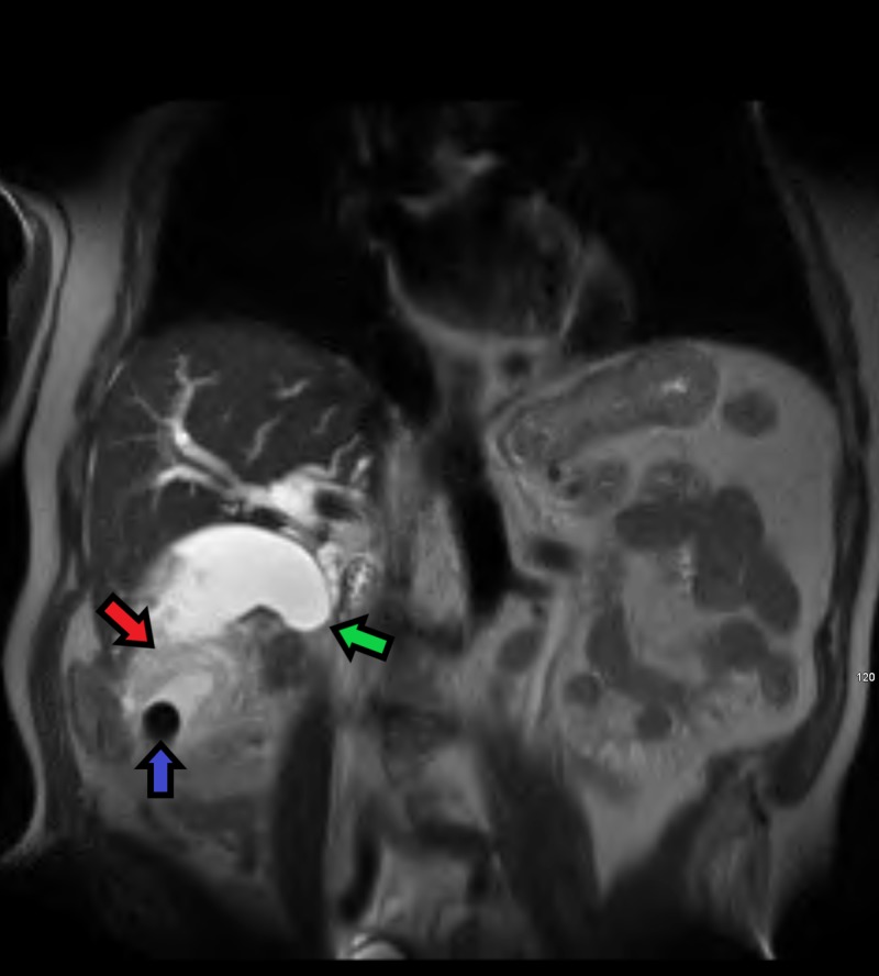
Figure 5. Coronal MRI T2 HASTE weighted image demonstrating a large mass (red arrow) surrounding a gallstone (blue arrow) in the distal portion of the gallbladder fundus. There is sharp tapering in the mid-to-distal common bile duct (green arrow) indicating obstruction secondary to infiltration by the mass.
MRI - Magnetic resonance image, HASTE - Half-Fourier Acquisition Single-shot Turbo Spin-Echo

