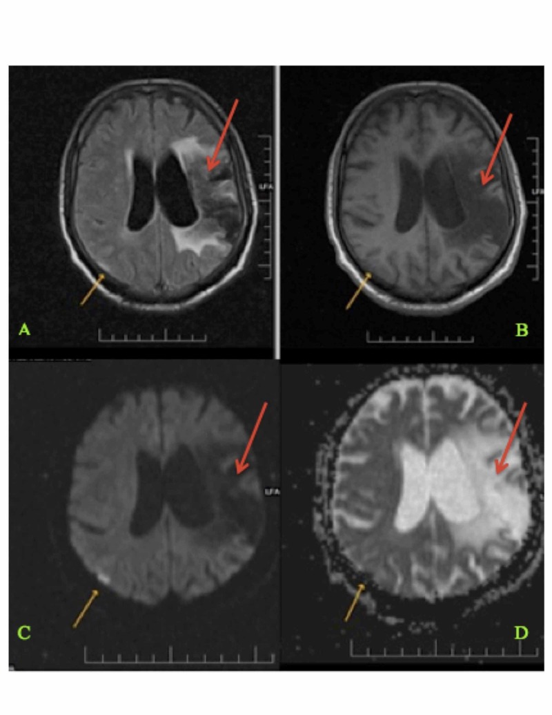
Figure 1. Magnetic Resonance Imaging (MRI) of the Brain Showing Current and Previous Infarction Sites (Panels A-D) .
Magnetic resonance imaging of the brain showing an acute right-sided parietal lobe infarct (shown with yellow arrows) and an old left-sided middle cerebral artery territory infarct (shown with red arrows).

