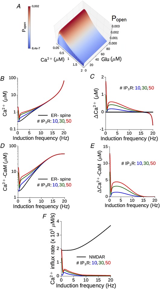
Figure 5. Augmented Ca2+–CaM response in a spine head in the presence of ER depends on the synaptic input rate and is suppressed at higher frequencies.

A, the steady state open probability of an IP3 receptor (P open) as a function of (constant) glutamate and Ca2+ concentrations. B, maximum Ca2+ level attained during persistent stimulation at different frequencies in the reference ER− spine (black) and equivalent ER+ spine with different numbers of IP3Rs (coloured curves). C, non‐monotonic dependence of the differential Ca2+ responses in the ER+ spine on the input rate. D and E, the corresponding results for CaM activation as a function of the input rate. F, the maximum calcium influx rate through NMDA receptors (black) and different numbers of IP3 receptors (coloured curves) in an ER+ spine plotted against the input rate. (All results for the model synapse with peak NMDAR‐mediated Ca2+ response ΔCaEPSP = 0.2 μM.) [Color figure can be viewed at wileyonlinelibrary.com]
