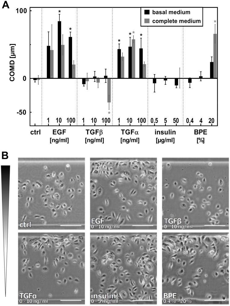
Fig 4. EGF, TGFα and BPE induce directed migration of nHEK.
A. nHEK cells were seeded in migration arenas coated with fibronectin. Gradients of EGF, TGFα, TGFβ, insulin, and BPE were established in the chemotaxis chamber, and the effect on cell migration was evaluated after 20 hours. The maximal concentration of each gradient is indicated in the graph. All gradients start from zero, with the exception of insulin and BPE, which were contained already at low concentrations in complete medium (0.4% BPE, 5 μg/ml insulin). In basal medium, the gradients of these factors also started from zero. Mean COMD ± SEM (n = 4); * indicate means significantly different from control (ANOVA followed by Dunnett’s test; p<0.05). Mean COMD ± SEM is also listed in a table in S5 Fig. B. Micrographs show cell distribution in the arenas at the end-point of the experiment (scale bars = 200 μm).

