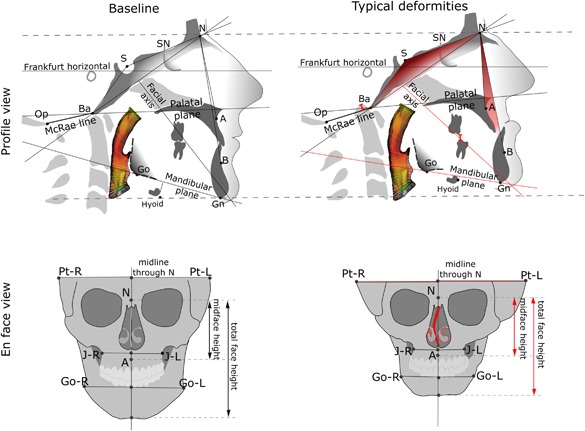
Figure 7.

Schematic illustration of the typical craniofacial deformation pattern observed in severe OI: (left) normal craniofacial parameters; (right) generalized trend of craniofacial deformities in severe OI. The more obtuse cranial base angle, the lower ANB angle, the reduced facial height, the overclosed mandible, the basilar invagination, the nasal septum deviation, and the cranial asymmetry in OI are indicated in red. Airway color mapping represents the variability of the cross‐sectional area.
