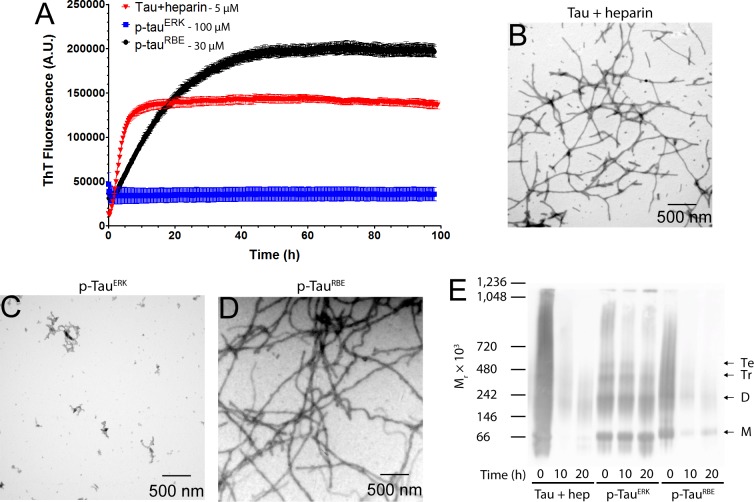
Figure 1.
Self-assembly of tau induced by heparin, p-tauERK and p-tau(S262A)RBE. (A) Time-dependent ThT fluorescence of 5 μM heparin-induced tau, 100 μM p-tauERK, or 30 μM p-tau(S262A)RBE. (B–D) Morphological examination by TEM at the end of the aggregation reaction (t = 100 h) of heparin-induced tau (B), p-tauERK (C) and p-tau(S262A)RBE (D). (E) Native PAGE analysis of 10 μM each of heparin (hep)-induced tau, p-tauERK, and p-tau(S262A)RBE at t = 0, 10, or 20 h of incubation under the same conditions. For heparin-induced tau, t = 0 is immediately after addition of heparin. Molecular weight markers are shown on the left. The mobilities of putative monomer (M), dimer (D), trimer (Tr), and tetramer (Te) are indicated on the right.

