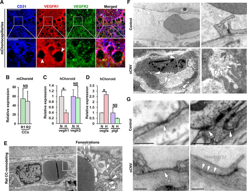
Figure 6.

Pathophysiological changes in the choriocapillaris (CCs) in adult zebrafish are recapitulated in mice, rats, and human wet age-related macular degeneration (AMD) patients. A, Confocal micrographs of CCs from adult C57/Bl6 mice (m) stained with anti-CD31 (blue), anti-VEGFR1 (red), and anti-VEGFR2 (green) antibodies. Boxed regions are shown in the magnified images below. White arrowheads indicate interstitial areas/extravascular columns. Size bars indicate 100 µm for the top row and 20 µm for the lower row. B, Quantifications of the percentage of the CD31+ endothelium that was costained with the VEGFR (R1) and VEGFR (R2) antibody in the experiment shown in A. n=3. C, Quantitative PCR analysis of Vegfr1 and Vegfr2 mRNA expression in primary human choroidal endothelial cells (hChoroid) in culture following 12 h of treatment with normal oxygen (normoxia, N) or 1% oxygen (hypoxia, H), normalized to the expression of TATA box-binding protein. n=3. *P<0.05. D, Quantitative PCR analysis of Vegfa or Plgf expression in primary human choroidal ECs (hChoroid) subjected to normoxia or hypoxia as in C. n=3. *P<0.05, ***P<0.001. E, Transmission electron micrographs of rat CC remodeling following adeno-associated virus-mediated VEGF (vascular endothelial growth factor)-A overexpression in the outer retina/subretinal space. Boxed region is shown in the magnified image to the right. White arrows point to endothelial luminal processes (ELPs), tight junctions (TJs), vesicles (V), endothelial thickness (ET), and fenestrations, as indicated. F, Transmission electron micrographs of human occult choroidal neovascularization (CNV) or non-CNV control CCs. Boxed regions are shown in magnified images to the right. White arrows point to ELPs and ET. Size bars indicate 2 μm or 1 μm, respectively. G, Transmission electron micrographs of details from human occult choroidal neovascularization (oCNV) or non-CNV control choriocapillaris. White arrows point to fenestrations (F) and TJs, as indicated. Size bars indicate 1 μm. NS indicates not significant.
