
Figure 2. IL-10–/– Mice Lacking PTPN2 in Myeloid Cells Develop More Severe Colitis.
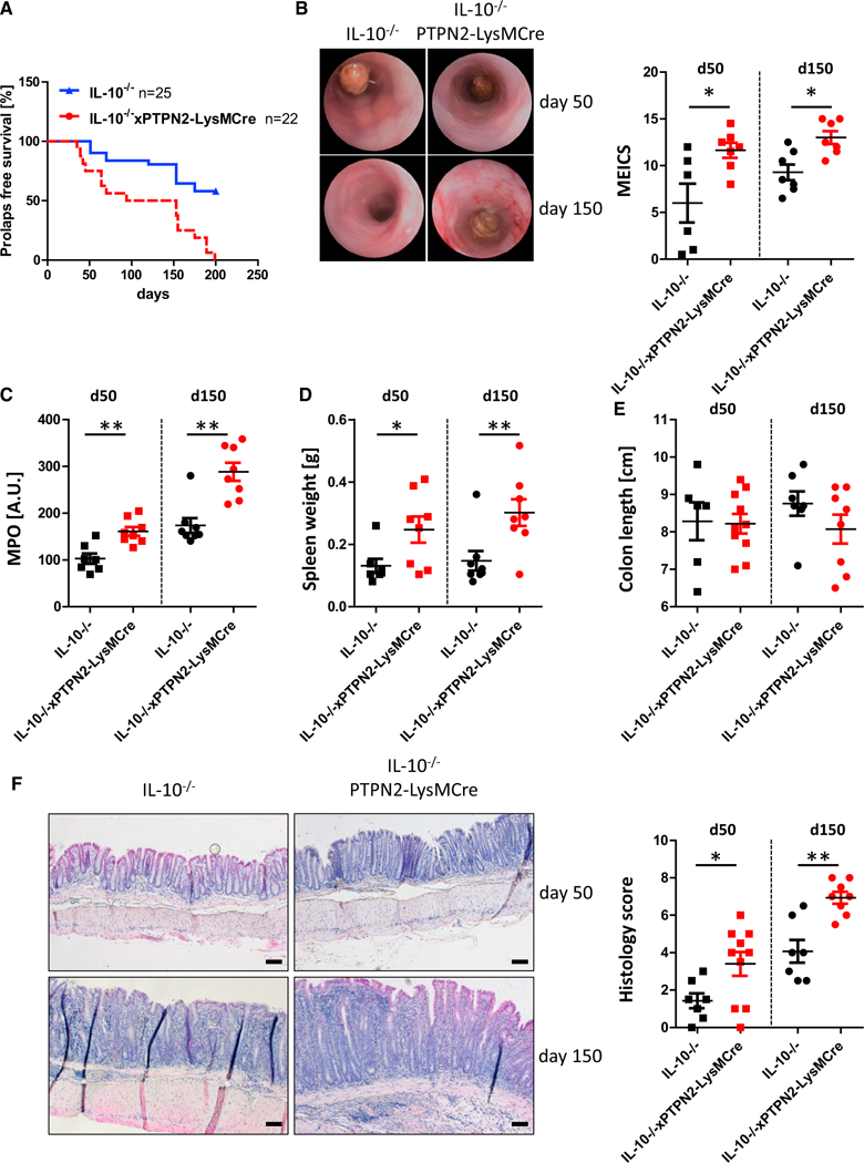
(A) IL-10–/– mice lacking PTPN2 in myeloid cells (IL-10xPTPN2-LysMCre) and their IL-10–/– littermates were monitored for 200 days for prolapse incidence. At 50 and 150 days, mice that did not develop prolapses were analyzed for colitis severity parameters.
(B–F) Shown are representative pictures from colonoscopy and respective statistical analysis (B), myeloperoxidase (MPO) activity (C), spleen weight (D), colon length (E), and representative pictures and scoring of epithelial damage and inflammatory infiltration in the terminal colon (F).
Depicted are results from seven or eight mice per group. Asterisks denote significant differences (*p < 0.05 and **p < 0.01; Mann-Whitney U test with Bonferroni correction). Scale bars represent 100 mm. Error bars represent means ± SD.