
Figure 6. Inhibition of IL-1 b Rescues PTPN2-LysMCre Mice from Increased Colitis.
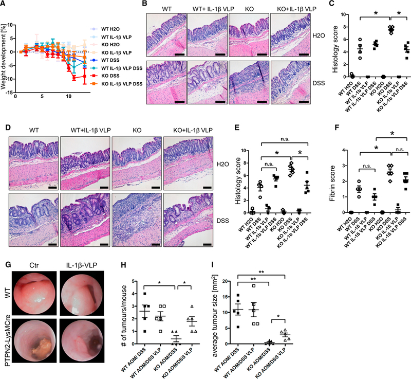
WT and PTPN72-LysMCre(KO) mice were immunized with IL-1β-coupled Qβ virus-like particles (IL-1β VLP) or with control Qβ virus-like particles 5,3, and 1 week prior to start of colitis/tumor induction.
(A–C) Weight development (A), representative pictures of H&E-stained pieces of the terminal colon (B), and results of histological scoring of intestinal pathology
(C) from mice with acute DSS colitis.
(D–F) Representative pictures from colonoscopy (D), statistical analysis of histopathology (E), and fibrin score from mice with chronic DSS colitis (F).
(G–I) Representative pictures of colonoscopy (G), observed tumor number in dissected colon (H), and average tumor size (I) in each mouse from mice subjected to AOM/DSS treatment.
Results are representative of one of two independent experiments with three to five mice per group. Asterisks denote statistical significance (*p < 0.05, **p < 0.01, and *** = p< 0.001; Mann-Whitney U test with Bonferroni correction). Scale bars represent 100 mm. Error bars represent means ± SD. See also Figures S8 and S9.