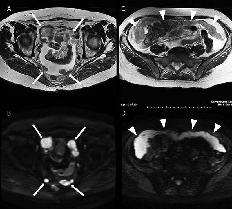
Figure 6.
Peritoneal metastatic nodular deposits (solid arrows) demonstrated on axial T2 (A) and diffusion-weighted imaging (B) sequences in the same patient. Omental cake (arrowheads) on axial T2: (C) intermediate signal: grey and prominent restricted diffusion on diffusion-weighted imaging; (D): high signal: bright.

