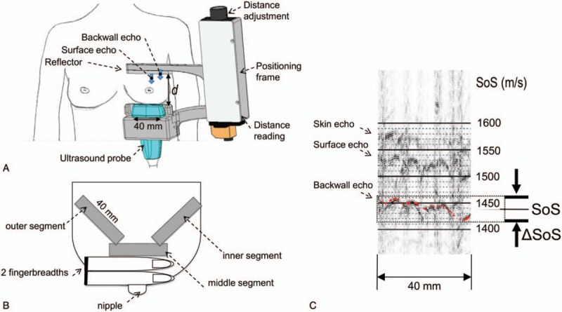
Figure 1.

Speed-of-Sound (SoS) ultrasound (US) examination setting. (A) View from ventral: breast with positioning frame, reflector, US probe and distance adjustment unit; (B) right breast viewed from above showing the three measured breast segments; (C) Annotation of average SoS value (1562 m/s) and SoS variation range (‘breast heterogeneity’) ΔSoS = 12 m/s in a dense breast segment. The back wall echo of the reflector is used as a timing reference. The automatic reading is displayed as an overlaid red line.
