Figure 2.

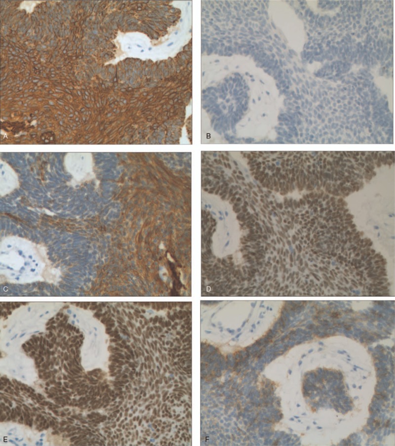
(a–f). Immunohistochemistry staining of ameloblastoma lung metastasis tissue (original magnification×200). The positive of CK(a), P40(d), P63(e), the weakly focally positive of CK5/6(c), CD56(f) and the negative of TTF-1(b).

(a–f). Immunohistochemistry staining of ameloblastoma lung metastasis tissue (original magnification×200). The positive of CK(a), P40(d), P63(e), the weakly focally positive of CK5/6(c), CD56(f) and the negative of TTF-1(b).