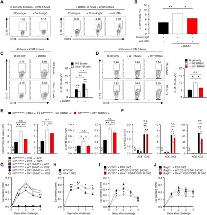
Fig. 6. MC-derived IL-5 is critical for the development of peripheral IL-10+ B cells to suppress CHS in mice.

(A and B) Splenic CD19+ B cells (1.5 × 10⁶ cells per well) were cocultured with an equal number of BMMCs with an anti–IL-5Rα mAb or isotype mAb for 43 hours and subsequently cultured with LPIM for 5 hours. Representative flow cytometry images (A) and histograms of the frequency of IL-10+ B cells (B) are shown. PE, phycoerythrin. (C and D) In (A) and (B), WT or Il5ra−/− splenic B cells were cocultured with equal numbers of WT or Il5v/v BMMCs as indicated. Representative flow cytometry images (left) and the frequency of IL-10+ B cells (right) are shown. (A to D) The results are expressed as representative images (A, C, and D) and mean ± SEM (C and D) from three independent experiments (triplicate for each experiment). **P < 0.01 by one-way ANOVA with post hoc Tukey’s test as indicated. (E and F) The histograms show the numbers of CD1dintCD5+ B cells and IL-10+ B cells in LNs (E) or IL-13+ ILC2s in LNs and ear tissues (F) from KitW-sh/W-sh mice with or without intravenous transfer of WT or Il5v/v BMMCs. (E and F) Data are expressed as the mean ± SEM from three independent experiments (n ≥ 3 per group for each experiment). *P < 0.05; **P < 0.01; n.s., not significant by one-way ANOVA with post hoc Tukey’s test. (G) The graphs show the ear thicknesses of KitW-sh/W-sh mice induced by OXZ with or without the reconstitution of WT or Il5v/v BMMCs. (H) The graphs show the ear thicknesses of WT or Il5ra−/− mice induced by OXZ. (I and J) The graphs show the ear thicknesses of Cd19Cre mice (I) or Rag2−/− mice (J) induced by OXZ with or without the transfer of WT or Il5ra−/− CD1dintCD5+ B cells. (G to J) Data are expressed as the mean ± SEM from two independent experiments (n ≥ 3 per group for each experiment). *P < 0.05, **P < 0.01 versus KitW-sh/W-sh mice (G), WT mice (H), Cd19Cre mice (I), or Rag2−/− mice (J) by Student’s t test.
