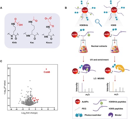
Fig. 1. Capture of the binding proteins of Khib.

(A) Structures of acetylation, succinylation, and 2-hydroxyisobutyrylation. (B) Workflow of the capture and enrichment of binding proteins of Khib by self-assembled multivalent photoaffinity peptide probes. UV, ultraviolet; AuNP, gold nanoparticles. (C) Volcano plot of enrichment result. The identified proteins with an abundance increase of more than twofold (log2[S/C] > 1) and P value less than 0.05 were marked red.
