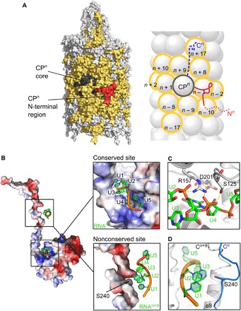
Fig. 2. Protein-protein and protein-RNA interaction networks in PVY.

(A) Surface (left) and schematic (right) representation of the CPn unit interacting with neighboring CP units (yellow). N-terminal CP region, red; CP core, dark gray; extended CP C-terminal region inside the filament, blue dashed line. N- and C-terminal regions in the scheme are depicted only for CPn. (B) Left: Electrostatic surface of CPn (red, negative; blue, positive) with two ssRNA interaction sites. Top right: Close-up view showing NUC1 to NUC5 nucleotides bound to the conserved site in the CPn core. Bottom right: Nucleotides binding to CPn+9 core also binding to Ser240 in CPn C terminus. (C) Conserved residues Ser125, Arg157, and Asp201 in the ssRNA binding site of the core. Electrostatic interactions are shown as dashed lines. For residues shown in sticks, protein/ssRNA: carbon, gray/green; oxygen, red; and nitrogen, blue; phosphorus, orange. (D) Nucleotides NUC1 to NUC3 binding to CPn+9 core facing Ser240 of CPn.
