Figure 5.
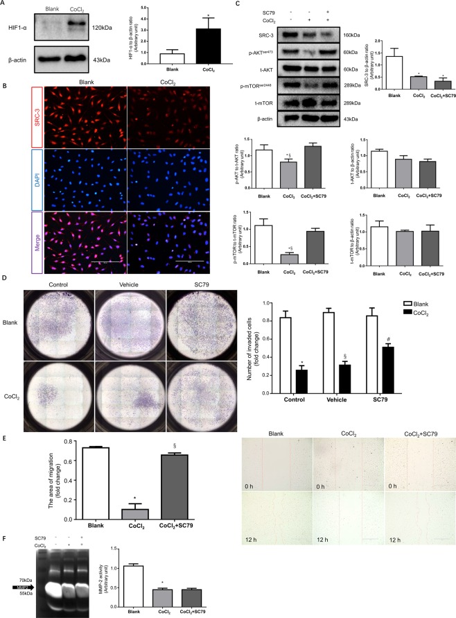
Hypoxia impairs the migration and invasion of HTR8/SVneo cells through inhibition of the SRC-3/AKT/mTOR signaling axis. After 48 h of 250 μM CoCl2 treatment, HTR8/SVneo cells were subjected to: (A) Western blot analysis of HIF1-α. (B) IF staining of SRC-3, scale bar: 200 μm. (C) Western blot analysis of SRC-3, AKT, p-AKT, mTOR, and p-mTOR levels in HTR8/SVneo cells in the presence of 250 μM CoCl2 and/or 20 μM SC79. The blank control was also included, n = 3, *p < 0.05 vs. Blank, §p < 0.05 vs. CoCl2 + SC79. (D) HTR8/SVneo cells subjected to Matrigel transwell assays in the presence of vehicle (0.1% DMSO), 250 μM CoCl2, and/or 20 μM SC79. Invaded cells were counted after 24 h. n = 3, *p < 0.001 vs. Blank control, §p < 0.001 vs. Blank vehicle, #p < 0.01 vs. CoCl2 control. (E) Wound-healing assays for HTR8/SVneo cells in the presence of 250 μM CoCl2 and/or 20 μM SC79. Images were taken at 0 h and after 12 h of treatment. The areas of migration were quantified in the bar graph. n = 3, *p < 0.001 vs. Blank, §p < 0.001 vs. CoCl2. (F) Gelatin zymography of MMP-2 activity in culture medium of HTR8/SVneo cells in the presence of 250 μM CoCl2 and/or 20 μM SC79, *p < 0.001 vs. Blank. All experiments were repeated three times.

