Figure 2.
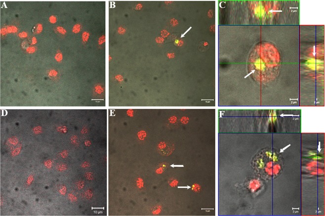
In vivo hemocytic test in Tenebrio molitor showing ND passage through the cuticle. Insect hemocytes after injection (B,C) and topical application (E,F) of FITC-NDs (at a dose of 2 ng or 5 µg of FITC-NDs per insect, respectively). Control beetles received physiological solution through injection (A) or topically (D). Regardless of the route of FITC-NDs administration to the beetles, they were visible inside the cytoplasm of hemocytes as yellow-green aggregates (arrows). Hemocyte nuclei were stained with propidium iodide solution. Scale bars: 10 µm (A,B,D,E) and 2 µm (C,F).

