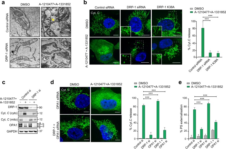
Fig. 5. DRP-1 regulates BH3 mimetic-induced cytochrome c release and apoptosis downstream of mitochondrial cristae remodelling.
a Electron microscopy of H1299 cells, transfected with DRP-1 siRNA for 72 h in the presence or absence of the indicated BH3 mimetics for 2 h. Breaks in the outer mitochondrial membrane are indicated by the yellow arrowhead. Scale bars = 10 nm. b H1299 cells were transfected with DRP-1 siRNA or GFP-DRP-1 K38A plasmid for 72 h, exposed to Z-VAD.fmk (30 µM) for 0.5 h, followed by a combination of A-1331852 (100 nM) and A-1210477 (10 µM) for 4 h and the extent of cytochrome c released from mitochondria assessed by confocal microscopy. The boxed regions in the images are enlarged to show mitochondrial structural changes in the indicated cells. The extent of cytochrome c release was quantified by counting at least 100 cells from three independent experiments. c Same as b, but the extent of cytochrome c release as well as OPA1 processing and the silencing efficiency of DRP-1 siRNA were analysed by western blotting. d H1299 cells were transfected with the indicated siRNAs for 72 h, treated as described in b and the extent of cytochrome c release assessed and quantified. The extent of cytochrome c release was quantified by counting at least 100 cells from three independent experiments. e Same as d but the cells were exposed to BH3 mimetics in the absence of Z-VAD.fmk and the extent of apoptosis assessed by PS externalisation from at least three independent experiments. All scale bars, unless indicated: 10 µm. Error bars = mean ± SEM. Statistical analysis was conducted by one-way ANOVA (***P ≤ 0.001)

