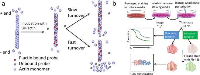
Figure 1.
Probing altered actin dynamics in live-cells based on an F-actin specific fluorogenic probe. (a) Schematic showing F-actin interaction with SiR-actin (SA), a cell permeable probe that fluoresces when bound to F-actin. Actin monomers are assembled/disassembled at either ends of the filament with different rates creating the fast growing “+” end and the slow growing “−” end. SA binds F-actin with high specificity, but upon removal from the filament due to actin turnover, the probe loses >100 fold in fluorescence. Probe binding is inversely correlated with actin turnover14. (b) SiR-Actin based Measurement of Actin Turnover (SMAT) involves prolonged live cell staining in the growth media with SA, removal of staining media, addition of cytoskeleton influencing cues and simultaneous time-lapse imaging. The loss of SA due to altered actin dynamics is quantified via the ratiometric decay of intensity in the image series captured during time “t” and normalized to the first timepoint (t0). Further, subtle phenotypic variations in the actin cytoskeleton are parsed via high content image analysis (HCIA), based on dual imaging of SA and F-actin specific fluorescent probe, phalloidin (ph-488).

