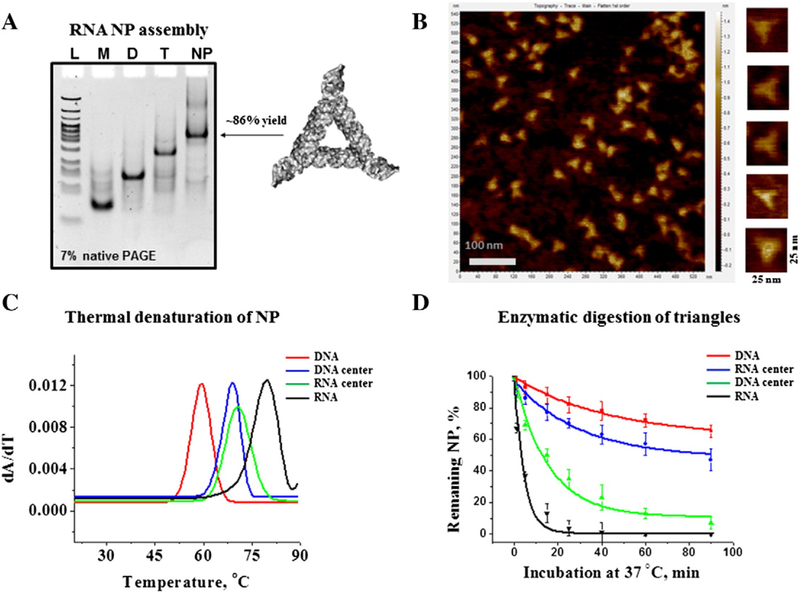
Figure 2.
RNA NP characterization. (A) Self-assembly properties of the triangle RNA were evaluated by 7% native PAGE in 0.5× TBM running buffer. Lane L is DNA ladder (Low Molecular Weight, New England Biolabs); Lanes M, D, and T are RNA monomer (rT2), dimer (rT2 and rT3), and trimer (rT2, rT3, and rT4) complexes, respectively. Lane NP (nanoparticle) contains fully assembled RNA triangle complex composed of all four RNA strands. The gel was stained in EtBr solution for the total RNA visualization. (B) RNA triangle AFM images acquired in air at ambient temperature. (C) Example of first derivatives of UV-melting curves of RNA, DNA and two hybrid RNA/DNA and DNA/RNA nanoparticles. The peaks correspond to Tm values of each individual NP. (D) Time dependent triangle NP (1 μM each) degradation profiles, obtained by incubation in 2% (v/v) fetal bovine serum at 37 °C demonstrating different decay rates. The data represent mean values of at least three independently repeated experiments.

