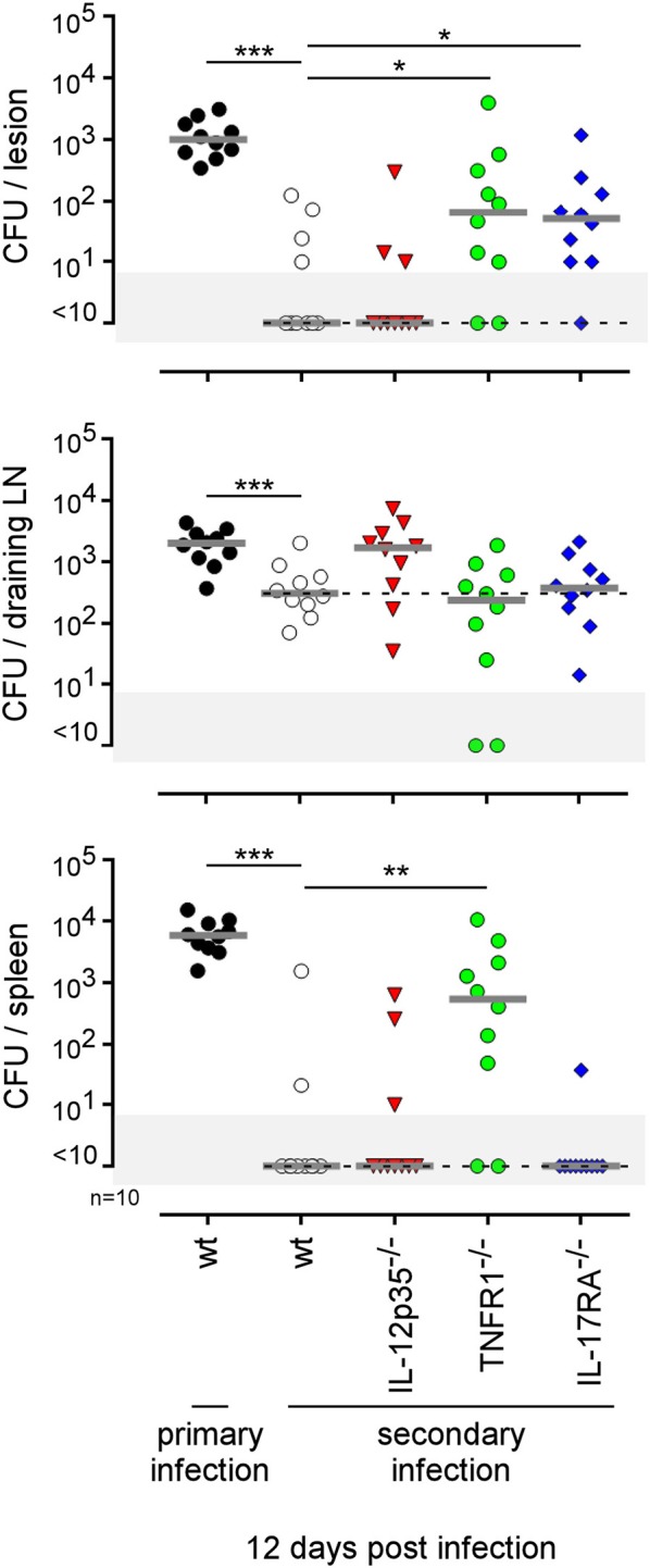
Figure 13.

Comparison of protection in wild-type and various deficient mice previously immunized by intradermal route with live B. melitensis. Wild-type, IL-12p35−/−, TNFR1−/−, IL-17RA−/− C57BL/6 mice were immunized intradermally (i.d.) with 2 × 10⁴ CFU of live wild-type B. melitensis and treated with antibiotics, as described in the Materials and Methods. Naive (primary infection group) and immunized (secondary infection group) mice were challenged i.d. with 2 × 10⁴ CFU of live mCherry-B. melitensis and sacrificed at 12 days post infection. The data represent the CFU count per organ. Gray bars represent the median. The significant differences between the indicated groups are marked with asterisks: *p < 0.1, **p < 0.01. These results are representative of three independent experiments. LN, lymph node; n, number.
