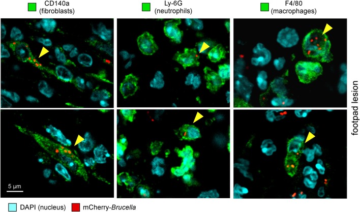
Figure 3.
Following intradermal infection with Brucella melitensis, fibroblasts, neutrophils, and macrophages were found to be infected in the footpad lesion. Wild-type C57BL/6 mice were infected intradermally with a dose of 107 CFU of mCherry-B. melitensis. Mice were sacrificed at 24 h post infection and the footpad lesions were collected and analyzed by confocal microscopy for the expression of mCherry and CD140a, LY6G, and F4/80 markers. The panels are color-coded with the text for DAPI, the antigen examined or mCherry-Brucella. Scale bar = 5 μm. Yellow arrowheads indicate the cells infected. The data are representative of two independent experiments.

