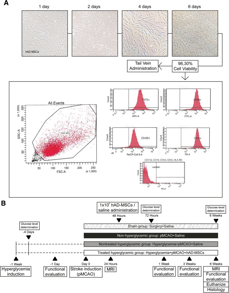
Fig. 1.
a Human AD-MSC cell culture protocol. Human AD-MSCs were thawed and cultured for their expansion on tissue culture flasks for 6 days. The phenotypic pattern of the cells was studied using flow cytometry, the positive expression of CD90, CD73, CD105 and CD44 (≥ 90%), and the lack of expression of CD11b, CD19, CD34, CD45, and HLA-DR were detected. Cell viability was studied and the cells were prepared for intravenous administration. b Experimental animal protocol. One week before stroke induction, hyperglycemia was induced in the rats with two intraperitoneal injections (nicotinamide and 15 min later streptozotocin). After 72 h and 6 weeks, blood glucose concentrations were determined. Rats were subjected to a cortical stroke by permanent middle cerebral artery occlusion and divided into groups. Forty-eight hours after surgery, 1 × 10⁶ hAD-MSCs or saline solution was administered. Functional evaluation was performed before surgery and at 1 week, 3 weeks, and 6 weeks post-stroke, and MRI was analyzed at 24 h and 6 weeks post-stroke. Six weeks post-stroke, the animals were euthanized and histological analyses were performed. Abbreviations: pMCAO: permanent middle cerebral artery occlusion; MRI: magnetic resonance imaging; hAD-MSCs: human adipose tissue-derived mesenchymal stem cells

