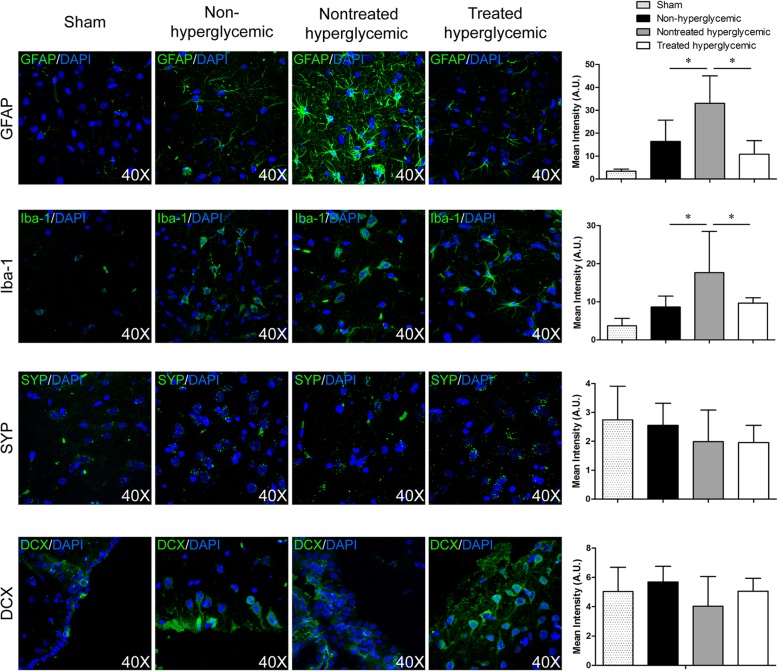
Fig. 4.
Brain repair markers related to glia and neurons at 6 weeks post-stroke by immunofluorescence. Representative images and quantification of immunofluorescence of GFAP, Iba-1, synaptophysin, and doublecortin. At 6 weeks post-stroke, the hyperglycemic rats showed significantly higher levels of GFAP and Iba-1 compared with the non-hyperglycemic and treated hyperglycemic groups. 4′,6-Diamidino-2-phenylindole (DAPI) was used for nuclear staining (3 animals per group, 4 sections in each animal per group). Data are shown as mean ± SD. *p < 0.05. Abbreviations: GFAP: glial fibrillary acidic protein; SYP: synaptophysin; DCX: doublecortin

