
Fig. 3.
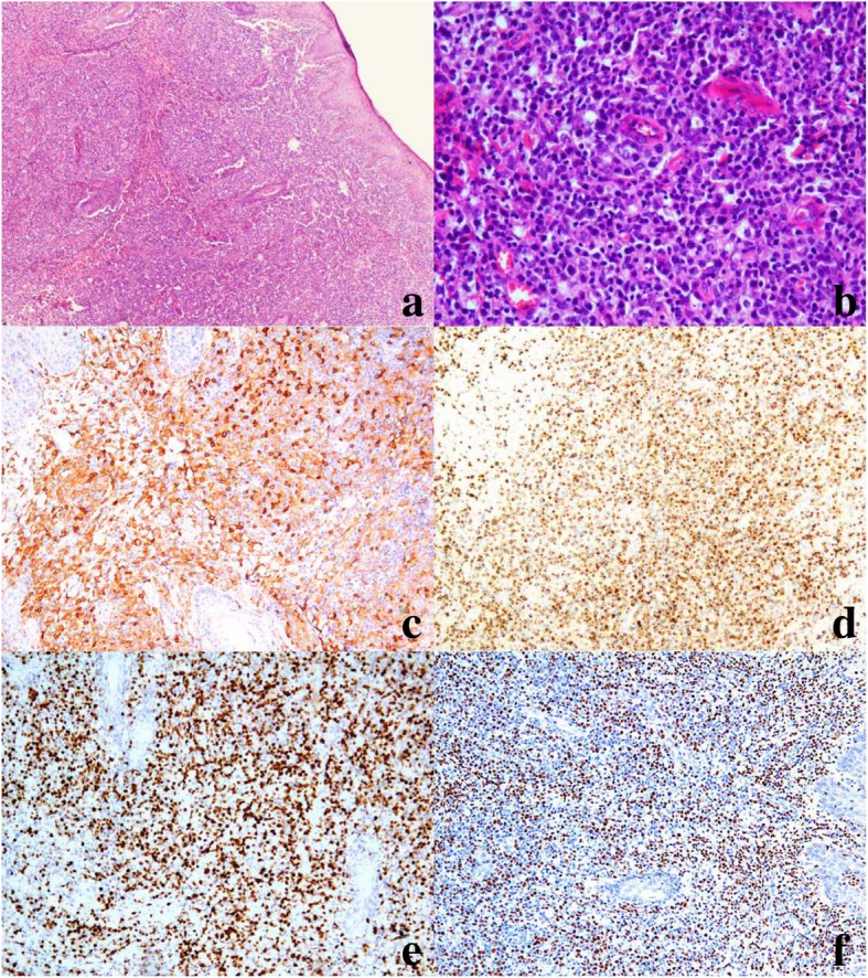
Morphology and immunophenotype of case 9, which progressed to systemic T-cell lymphoma. a Skin biopsy shows a diffuse, atypical lymphocyte infiltrate, with a marked epidermis and subcutaneous distribution (hematoxylin and eosin stain [H&E], 40×). b Skin biopsy shows medium- to large-sized lymphocytes, with enlarged, oval and pleomorphic nuclei (H&E, 200×). c Strong staining of CD4+ cells (IHC, 200×). d GranB Positive cells (IHC 100×). e Ki67 expression is high in 70% of tumor cells (IHC 100×). f In situ hybridization shows that neoplastic cells are positive for EBV-encoded RNA (100×)