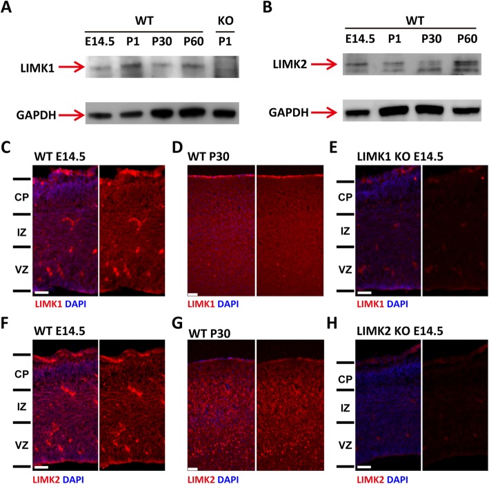
Fig. 1.
Expression of LIMK1 and LIMK2 in developing mouse neocortex. a, b Western blots showing LIMK1 (a) and LIMK2 (b) expression in mouse neocortex at various developmental stages. (c, d, e) Coronal brain sections co-stained with anti-LIMK1 and DAPI showing the expression of LIMK1 in WT E14.5 (c) and WT P30 (d), but not in LIMK1 KO E14.5 (e). (f, g, h) Coronal brain sections co-stained with anti-LIMK2 and DAPI on coronal brain sections of WT E14.5 (f) and WT P30 (g), but not in LIMK2 KO E14.5 (h). Note some remaining staining signals in LIMK1 and LIMK2 KO sections, suggesting that the antibodies might be non-specific. CP, cortical plate; IZ, intermediate zone; VZ, ventricular zone. Scale bars, 50 μm

