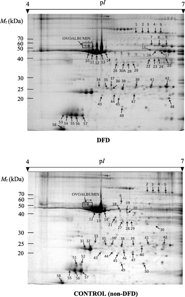
Fig. 1.

Representative 2-DE gel profiles of DFD (above) and control (below) meat samples from the LT bovine muscle stained with Pro-Q Diamond and subsequently with SYPRO Ruby. Phosphoprotein spots with statistically significant qualitative (presence/absence) and quantitative (changes in intensity) differential phosphorylation are marked and numbered. Numbered spots were excised from gels for phosphoprotein identification by MALDI-TOF and MALDI-TOF/TOF MS.
