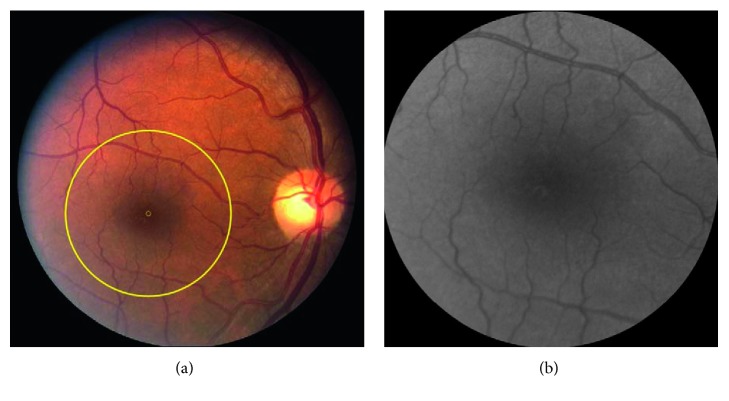
Figure 1.
(a) Example of a color retinal image acquired in a nondiabetic control subject. A circular region of interest (ROI) with a diameter of 3.6 mm centered on the fovea and outlined by a yellow circle was selected for discrimination analysis. The small yellow circle in the center of the large circle shows the center of the fovea. (b) Converted grayscale images of the ROI in the same subject.

